MED0001186584 This website contains imagery which is only suitable for audiences 18+. All procedures contain risks. Surgical risks read more here. Non-surgical risks read more here.
Doctor-performed removal of raised moles, skin tags, seborrheic keratosis, DPN and syringomas — using the Ellman Surgitron 4.0 MHz radiofrequency unit. Critical assessment with dermatoscopy comes first, every single time. Available at both Melbourne clinics: The DOC West (Hoppers Crossing) and The DOC East (Ashburton), with every consultation and treatment performed personally by Dr Ed Omarjee.
Important: every lesion must be examined in person, with dermatoscopy, before any treatment is recommended. Radiofrequency removes a lesion by vaporising it, so no tissue remains to send to a laboratory. Assessment always comes first, and cosmetic removal is only offered where a lesion is confidently benign. This document provides general information only and does not replace an individual medical consultation. All procedures carry risks.
Recent results — radiofrequency lesion removal
Before & after, from both clinics
Genuine patient results from both The DOC West and The DOC East, photographed on our standardised clinical imaging system and published with written consent. The strip below moves through recent cases; browse the full collection in the gallery.
DISCLAIMER: Outcomes shown are only relevant for the individual patient and do not necessarily reflect the results other patients may experience, as results may vary due to many factors including genetics, skin type and healing. Some images may have patients’ tattoos, jewellery or other identifiable items blurred to protect patient identities.
Non-negotiable first step
Why the dermatoscope decides everything
Radiofrequency removes a lesion by vaporising it. That is excellent for cosmetic outcome — and it is exactly why the lesion must be confidently benign before it is treated. There is no second chance to examine tissue that no longer exists.
What a dermatoscope actually is
A dermatoscope is a handheld optical instrument: a high-quality lens giving around ×10 magnification combined with a controlled, polarised light source. Ordinary light bounces off the skin surface; polarising filters cancel that glare, so we see past the surface into the epidermis and upper dermis, where the architecture of a lesion is written.
Structures invisible to the naked eye become obvious: pigment networks, globules, streaks, vessel patterns, keratin plugs, blue-white veils. It is the difference between “that looks like a mole” and “that has the architecture of a benign intradermal naevus.”
Assessment is systematic, not a glance. Each lesion is examined for symmetry of structure, pigment distribution, vessel pattern, border and colour — and your history and any change you have noticed all feed into the decision. Dermatoscopy is non-invasive and painless, and it is the step that decides whether cosmetic removal is appropriate.
Dr Ed Omarjee assessing a lesion under dermatoscopy at The DOC. Right: the handheld polarised dermatoscope itself, giving around ×10 magnification. Every lesion is examined this way before any treatment is recommended.
What we look for
What we look for, and what it tells us
A simplified guide. In practice these features are weighed together, never in isolation, alongside your history and any change you have noticed.
Reassuring
Milia-like cysts and comedo openings — the classic signature of a seborrheic keratosis.
Comma-shaped vessels — short, evenly arranged curves, typical of a benign intradermal naevus.
Regular pigment network — an even honeycomb mesh that fades softly at the edges.
Symmetry of structure — the same architecture throughout the lesion.
Not reassuring
Arborising vessels — branching, tree-like vessels in sharp focus; a recognised feature of basal cell carcinoma.
Blue-white veil — a structureless blue-white area requiring excision and histopathology.
Asymmetry of structure — different architecture in different parts of the same lesion.
Disordered or abruptly ending network — thickened, irregular or sharply cut-off pigment.
Three possible outcomes
Three possible outcomes, and only one of them is removal
Outcome A
Confidently benign
The lesion’s dermatoscopic architecture matches a known benign pattern. Cosmetic radiofrequency removal can proceed, usually the same day if you are happy with the plan, the risks and the quote.
Outcome B
Uncertain
Features are not fully typical. We photograph and monitor over an interval, or take a diagnostic biopsy first. Nothing is vaporised while a question remains.
Outcome C
Suspicious
Features of concern. The lesion is surgically excised with margins and sent for histopathology, or you are referred appropriately. Cosmetic RF is not offered.
This is the single most important thing on this page. Radiofrequency destroys the tissue it removes, so no specimen goes to the laboratory. Any practitioner who offers to “zap off” a pigmented lesion without examining it under dermatoscopy is removing your ability to have that lesion diagnosed.
If your lesion is pigmented, has changed, is new after the age of 40, itches, bleeds, or simply looks different to your others, say so at your consultation. It changes the plan, and that is exactly as it should be.
Clinical imaging
Standardised clinical photography at both clinics
You cannot honestly judge a result against a memory. Both The DOC West and The DOC East are equipped with a dedicated, professionally installed clinical imaging system, so your before and after images are genuinely comparable.
Our dedicated photography room at The DOC East, Ashburton. Fixed lighting and marked floor positions for face and body shots mean every image is captured from the same spot, under the same light.
The camera is pre-calibrated and tethered directly to the imaging database, so settings never drift between visits and images file automatically to your record.
Why standardisation matters
Most before and after photographs you see online are not comparable. Change the lighting, the angle, the distance, the camera or the expression, and you change the apparent result, without a single thing having changed on the patient’s skin. That is not evidence; it is marketing.
Our clinics use a system from Clinical Imaging Australia, developed by Woodrow Wilson and used by many of Australia’s leading plastic surgery, cosmetic and dermatology practices. It combines a pre-calibrated camera and fixed studio lighting with software that manages the image database, so every photograph is taken under identical conditions.
Because the lighting is dedicated and controlled, the time of day and changes in ambient room light have no effect on your images. The result is that a photograph taken at your consultation and one taken at review three months later can be placed side by side and meaningfully compared, by Dr Ed Omarjee, and by you.
What the system gives us
Lighting
Fixed, calibrated
Angle & distance
Reproducible
Ambient light
No effect on images
Skin filters
Melanin & haemoglobin
Depth visualised
To roughly 2 mm
Storage
Australian data centres
Access
Two-factor authentication
Digital skin filters
The system supports multispectral views that isolate melanin and haemoglobin within roughly the first 2 mm of skin. Alongside handheld dermatoscopy, this helps map pigment distribution and vascular patterns, useful both for planning a removal and for documenting how pigmentation settles afterwards, particularly relevant in DPN and in richly pigmented skin.
Your images, and your privacy
Clinical photography forms part of your medical record. Images are stored securely behind two-factor authentication on Australian-based data centres, consistent with Privacy Act requirements for medical data.
Photographs taken for your medical record are never published without your separate, specific, written consent. Consenting to clinical photography for your record is not consent to appear in our gallery, on our website or on social media. Those are distinct decisions, and you are free to decline either. This approach reflects AHPRA’s advertising guidelines and TGA requirements around the use of before and after imagery.
Lesion atlas
What we remove, and what each one actually is
Benign raised lesions are not all the same thing. They arise from different cells, behave differently, and respond differently to treatment. Here is the practical guide to the ones we see most often.
Intradermal naevusThe classic raised mole
The most common raised mole in adults. Nests of naevus cells sit entirely within the dermis, pushing the skin upward into a soft, dome-shaped bump. Often skin-coloured, tan or light brown rather than dark, as many have lost pigment over decades. Frequently found on the face and neck, sometimes with a hair growing through.
People ask about these because they catch on razors, glasses and jewellery, or simply because they cast a shadow in photographs.
RF suitability: Excellent. The dome shape is ideally suited to being levelled to the surrounding skin plane.
Compound naevusRaised centre, flat pigmented edge
Naevus cells sit at both the junction of the epidermis and dermis and deeper within the dermis. Clinically this reads as a raised central portion with a flatter pigmented halo around it. Usually mid-brown and evenly coloured.
Assessment matters more here than with a purely intradermal naevus, because the junctional component is pigmented and needs dermatoscopic evaluation of its network.
RF suitability: Good for the raised portion once confirmed benign. The flat pigmented edge may persist, and this is discussed honestly beforehand so expectations are accurate.
Skin tagsAcrochordon, fibroepithelial polyp
Soft, skin-coloured or slightly darker outgrowths attached by a narrow stalk. They favour areas of friction: neck, underarms, eyelids, under the bust, groin. They are entirely benign, but they snag, they multiply, and they are frequently the thing people most want gone.
Numbers often increase with age, weight gain, pregnancy, and in insulin resistance. If you have developed many quite suddenly, it is reasonable to have your metabolic health checked with your GP.
RF suitability: Excellent, and fast. Many can be treated in a single sitting, which makes the additional-lesion pricing particularly relevant.
Extremely common from middle age onward. They have a characteristic “stuck-on” appearance, as though a piece of wax or bark has been pressed onto the skin, with a rough or warty surface and colours ranging from pale tan to almost black. Under dermatoscopy they show milia-like cysts and comedo-like openings, a very satisfying, reassuring pattern.
They are harmless, but they are the lesion most often mistaken by patients for something sinister, precisely because they can be dark and irregular.
RF suitability: Very good. Because they are superficial, they can usually be removed with minimal depth, which favours a flat result.
Dermatosis papulosa nigra (DPN)Multiple small dark papules
A variant of seborrheic keratosis seen predominantly in people with richly pigmented skin (Fitzpatrick types IV–VI), often with a strong family history. Presents as multiple small, dark brown to black, smooth raised papules across the cheeks, temples, forehead and neck. They typically begin in adolescence or early adulthood and slowly increase in number.
DPN is entirely benign. It is treated for appearance only, and it is one of the most rewarding treatments we do, because clearing dozens of lesions transforms how skin reads at conversational distance.
RF suitability: Very good, with an important caveat. Pigmented skin carries a real risk of post-inflammatory hyperpigmentation, so we use conservative settings, often treat a small test area first, and take a staged approach across sessions.
SyringomasBenign eccrine sweat duct tumours
Small, firm, skin-coloured to slightly yellow papules, typically 1–3 mm, clustered symmetrically under and around the eyes, though they also occur on the upper cheeks, forehead, chest and neck. They arise from eccrine sweat ducts, are more common in women, often begin around puberty, and frequently run in families.
They are completely benign and do not resolve on their own. Because they sit in a delicate area with thin skin, they demand a conservative, patient approach rather than aggressive treatment.
RF suitability: Good, but they are one of the harder lesions to treat well. They extend deeper than they appear, recurrence is genuinely common, and multiple sessions are usually needed. Treating too deeply near the eye risks a worse result than the syringomas themselves, so we treat lightly and repeat.
Sebaceous hyperplasiaEnlarged oil glands
Soft, yellowish papules with a characteristic central dimple or umbilication, most often on the forehead, nose and cheeks. They represent enlarged, overgrown sebaceous glands rather than any kind of growth, and become more common with age and in people with oilier skin.
Their key clinical importance is that they can mimic early basal cell carcinoma to the naked eye, which is precisely why dermatoscopy, where they show a distinctive crown of vessels around a central core, matters before anything is treated.
RF suitability: Very good. Fine, superficial treatment flattens them neatly. Recurrence over years is possible because the underlying gland remains.
Cherry angiomasCampbell de Morgan spots
Bright red to purple domed papules made of a small cluster of dilated capillaries. They appear from around the thirties onward, mostly on the trunk, and increase in number with age. Entirely benign, though they can bleed briskly if they are caught or scratched.
RF suitability: Excellent, and this is where radiofrequency shines, because the same energy that removes the lesion also seals the vessels feeding it.
Fibrous papule of the faceAngiofibroma
A single firm, dome-shaped, skin-coloured papule, classically on or beside the nose. Small, usually under 5 mm, and often mistaken for a persistent pimple that never came to a head, or for a mole. Benign, but worth confirming, since it can resemble a basal cell carcinoma or a naevus clinically.
MiliaKeratin-filled cysts
Firm, pearly white 1–2 mm bumps sitting just beneath the surface, most commonly around the eyes and on the cheeks. They are tiny cysts containing trapped keratin, not blocked oil, and not treatable by squeezing.
RF suitability: Straightforward. Often the fastest treatment we do: the cyst is opened and the contents released with minimal energy.
Viral wartsVerruca vulgaris
Rough, raised lesions caused by human papillomavirus, with a firm surface and often tiny dark dots representing thrombosed capillaries, a useful dermatoscopic giveaway. Common on hands, fingers and feet, and contagious.
RF suitability: Effective. Because warts are viral, recurrence is possible if virus persists in surrounding skin. This differs from the other lesions on this page and is discussed upfront.
XanthelasmaCholesterol deposits around the eyes
Soft yellowish plaques on the eyelids, particularly near the inner corners, caused by lipid deposition in the skin. Cosmetically bothersome and often gradually enlarging.
Important: xanthelasma can be a marker of abnormal blood lipids or increased cardiovascular risk. We will recommend lipid testing with your GP alongside any cosmetic treatment.
RF suitability: Possible, in a special facial area, with careful technique. Recurrence is common if the underlying lipid problem is not addressed.
Not on this list? This atlas covers what we see most often, not everything that exists. Dermatofibromas, epidermoid cysts, lipomas, keloids and a range of other lesions each need their own approach, and some are better served by surgical excision than by radiofrequency.
Dr Ed Omarjee has a particular clinical interest in keloid scarring, especially ear keloids. If your concern is a thickened, growing scar rather than a mole, that is a different treatment pathway. Please mention it when booking, or read about ear keloid removal.
The instrument
The Ellman Surgitron, and why 4.0 MHz matters
Most people never ask what device is being used on their face. They should. The physics of the instrument is one of the largest single factors in how the treated skin will look in six months.
4.0 MHz
Operating frequency
High-frequency radiowaves, in the broadcast radio range. Not a heated tip, and not a laser.
The tissue
Heat source
Heat is generated inside the cells themselves by their resistance to the wave, not transferred in from outside.
Stays cool
Electrode
The fine wire tip does not glow and does not burn. It guides energy; it does not deliver heat by contact.
Minimised
Lateral spread
Higher frequency concentrates the effect, narrowing the zone of collateral thermal damage either side of the cut.
The physics, in plain English
A radiofrequency unit passes a high-frequency electrical waveform from a fine electrode, through your tissue, to a return antenna plate positioned nearby. As that wave meets the natural resistance, the impedance, of the tissue in its path, energy is deposited within the cells themselves.
The water inside those cells heats extremely rapidly and turns to steam. The cells effectively burst apart, and the tissue parts in front of the electrode. This process is called cellular volatilisation. Nothing is being crushed, torn, scraped or burned by contact: the tissue is being separated from within, at cellular scale.
Two consequences follow, and both matter for your result. First, the electrode itself never becomes hot, so there is no branding of adjacent skin. Second, because the doctor is not pushing against tissue with a blade, there is no mechanical trauma to the wound edges.
A treatment room at The DOC, equipped with the Ellman Surgitron radiofrequency unit alongside our laser platforms. Mode and power are selected for each individual lesion.
Waveform selection
Each waveform trades cutting precision against bleeding control. The choice is made lesion by lesion: more cut for cosmetic precision, more coagulation only where bleeding demands it.
Pure cut
Least lateral heat. Used where cosmetic precision matters most.
Cut with coagulation
The everyday setting. Removes tissue while controlling small vessels.
Coagulation
Maximum bleeding control, more lateral heat. Used sparingly.
Why frequency changes the scar
Here is the part that actually determines the cosmetic outcome. Any electrosurgical device produces some heat spreading sideways from the point of contact. That band of collateral damage is called the zone of lateral thermal damage, and it is the tissue that has been cooked but not removed.
Damaged-but-surviving tissue is what the body must repair. More of it means more inflammation, more collagen laid down in repair, and a greater chance of a thickened, textured or discoloured mark. Less of it means a flatter, quieter heal.
As operating frequency rises, that zone narrows. At roughly 4.0 MHz the unit sits in the practical sweet spot: high enough for a genuinely fine, low-damage cut, while still delivering the haemostasis needed to keep the field clear. That combination is exactly what cosmetic facial work demands.
Safety, in short
Implanted electrical devices are the key contraindication. Radiofrequency is usually not used where you have a pacemaker, implantable defibrillator, nerve or spinal cord stimulator, or cochlear implant. Dr Ed Omarjee will usually consider alternatives in these situations, most often laser-based.
Routine precautions every case: the return antenna is positioned correctly, alcohol skin preparation is allowed to dry fully before activation, and the smoke plume is actively evacuated. Note that histology is not possible after RF, which is why assessment comes first.
How it differs
Radiofrequency is not diathermy, and not cautery
These terms get used interchangeably in clinics and online. They describe genuinely different machines with different effects on tissue.
Property
Ellman RF radiosurgery
Our device
Conventional diathermy / hyfrecator
Electrocautery
How heat is made
Ellman RF radiosurgery
Generated inside the cells by tissue impedance
Conventional diathermy / hyfrecator
Current through tissue, lower frequency
Electrocautery
A hot wire. Heat conducted in by contact
Typical frequency
Ellman RF radiosurgery
Around 4.0 MHz
Conventional diathermy / hyfrecator
Commonly well below 4 MHz
Electrocautery
None. No current enters the patient
Electrode temperature
Ellman RF radiosurgery
Stays cool
Conventional diathermy / hyfrecator
Warms with use
Electrocautery
Glowing hot by design
Lateral thermal damage
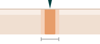
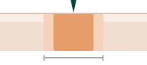
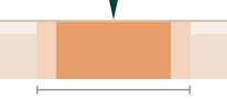
Cross-section through the treated site. The solid band is dense thermal damage, the pale band its falloff.
Ellman RF radiosurgery
Narrowest of the three
Conventional diathermy / hyfrecator
Wider
Electrocautery
Widest
Charring / eschar
Ellman RF radiosurgery
Minimal
Conventional diathermy / hyfrecator
Moderate
Electrocautery
Marked
Cosmetic control
Ellman RF radiosurgery
Fine, layer-by-layer sculpting
Conventional diathermy / hyfrecator
Less refined
Electrocautery
Blunt. Haemostasis, not contouring
Best used for
Ellman RF radiosurgery
Cosmetic facial lesion removal
Conventional diathermy / hyfrecator
General minor procedures, vessel work
Electrocautery
Stopping bleeding, field surgery
Comparative characteristics of the technologies. Outcomes in practice depend at least as much on operator technique, lesion selection and aftercare as on the device. No instrument removes the need for careful hands and correct patient selection.
Your appointment
What actually happens on the day
Most people are in and out inside an hour, including the consultation. Here is the sequence, so nothing is a surprise.
Radiofrequency removal in progress. The electrode tip is hair-fine and stays cool. The lesion is reduced in fine layers under local anaesthetic, with the surrounding skin supported and the profile checked against the skin plane as we go.
1Consultation and dermatoscopic examination
Dr Ed Omarjee examines each lesion you are concerned about, and looks at your skin more broadly. Your history, any changes you have noticed, your skin type and your scarring history all get discussed. You will be told plainly which lesions are suitable, which are not, and why.
2Standardised clinical photography, plan and quote
Baseline clinical photographs are taken with your consent using our dedicated Clinical Imaging Australia system, installed at both clinics, the same standardised setup used by leading plastic surgery and dermatology practices. You then receive a firm quote before anything proceeds. If you would prefer to think it over, that is completely fine.
3Cleaning and local anaesthetic
The area is cleaned and a small injection of local anaesthetic is placed. This is the only genuinely uncomfortable part, a brief sting lasting seconds. We use a fine needle and take our time. Once it is working, the area is completely numb.
4Radiofrequency removal
The lesion is reduced in fine layers, with the doctor repeatedly checking the profile against the surrounding skin plane. Bleeding is controlled as we go. You will hear a faint sound and may smell the treatment, and both are normal. You will not feel pain. On rare occasions, particularly in looser or more flaccid skin, a suture may be needed to close the site neatly.
5Hydrocolloid dressing and written aftercare
The treated area is covered with a small specialised hydrocolloid dressing, which stays in place for a few weeks to support healing in a moist environment. You leave with written instructions rather than something to try to remember. Most people drive themselves home and return to work the same day.
6Review
A review is organised at an appropriate interval, weeks to months after the procedure, if anything is concerning you. We check healing, assess the final result, and address anything that needs it, including early management if your skin is showing any tendency to thicken or pigment.
Recovery
Healing, honestly
Removing tissue always leaves a mark while the skin repairs. The goal is a flat, pale, unremarkable result, not an invisible one on day three.
Days 0–2
A small hydrocolloid dressing is applied and stays on. Mild redness and slight swelling are expected underneath.
Days 3–10
A crust forms and firms beneath the dressing. Let it lift on its own: picking it is the single most reliable way to worsen a scar.
Weeks 2–6
The dressing comes off after a few weeks, revealing fresh pink skin. It will look pinker than surrounding skin. Daily SPF 50+ is essential from here.
Months 2–12
The pink fades and the area blends. Facial skin usually settles fastest; the chest, shoulders and back take longest.
Scarring, pigment, and being straight with you
Radiofrequency is chosen because it tends to heal flatter and less conspicuously than a scalpel excision, which leaves a linear scar and needs sutures. It reduces risk. It does not abolish it.
Three specific things influence how you heal, and we assess all three before treating.
Your skin tone
Richly pigmented skin (Fitzpatrick IV–VI) carries a genuine risk of post-inflammatory hyperpigmentation, a temporary darkening at the treated site that can take months to settle. This is why we treat DPN conservatively and often in stages.
Your scarring history
If you or your family form keloid or hypertrophic scars, that changes the plan. Some sites, the chest, shoulders, upper back and earlobes, are higher risk regardless of history.
The lesion itself
Larger and deeper lesions need more tissue removed, and more removal means a more visible mark. A 2 mm skin tag and a 9 mm dome-shaped naevus do not heal the same way, and we will say so.
Aftercare essentials
Hydrocolloid dressing
In place a few weeks
Keep dry
First 24–48 hours
Ointment
As directed, keep moist
Picking the crust
Never
Makeup
Once fully sealed
Swimming / spa
Wait until healed
Sun protection
SPF 50+ daily, 6+ months
Heavy exercise
48 hours on the face
A review is organised at an appropriate interval, weeks to months post-procedure, if anything is concerning you. Contact the clinic sooner if you develop increasing pain, spreading redness, heat or discharge, as these are not part of normal healing.
Pregnancy & breastfeeding
If you are pregnant or breastfeeding
Short version: purely cosmetic removal is best deferred. But if a benign lesion is genuinely bothering you, it can be treated during pregnancy. And assessment is never delayed, at any stage.
Symptomatic benign lesions can be treated. If a confirmed benign lesion is causing you real trouble, a skin tag catching on clothing, jewellery or a bra strap, one that is repeatedly irritated, inflamed, snagged or bleeding, or a lesion in a spot that rubs constantly, there is no reason you should put up with it for nine months. Dr Ed Omarjee will treat these during pregnancy where it is warranted, after dermatoscopic assessment confirms the lesion is benign.
We limit the number of lesions per session. This is the key modification. Rather than clearing everything in one long appointment, we deliberately restrict how many lesions are treated at a time, specifically to keep the total dose of local anaesthetic as low as possible. If you have several bothersome lesions, they are staged across separate visits. It is a small inconvenience that keeps the anaesthetic exposure minimal, and it is the sensible way to do this.
Purely cosmetic removal is usually deferred. If a lesion is not bothering you physically and you simply do not like the look of it, our advice is to wait until after pregnancy and breastfeeding. There is no clinical urgency, and there are three good reasons to hold off.
Reason one
Pregnancy changes your skin, which changes the assessment
Hormonal shifts commonly cause existing moles to darken or enlarge, and can bring on melasma and generalised pigmentation. These physiological changes make dermatoscopic interpretation harder and less definitive. Waiting until your skin settles gives a cleaner assessment.
Reason two
Healing and pigmentation behave differently
Raised oestrogen and increased melanocyte activity mean a higher likelihood of post-inflammatory hyperpigmentation after any procedure that inflames the skin. A removal that would heal invisibly at another time can leave a lingering brown mark.
Reason three
Pregnancy skin tags often resolve on their own
New skin tags are very common in pregnancy, particularly on the neck, under the bust and in the underarms. A meaningful proportion shrink or disappear in the months after delivery. If they are not troubling you, waiting may mean you never need to pay to remove them at all.
About local anaesthetic. Lignocaine (lidocaine) is widely used during pregnancy where a procedure is warranted, dental treatment being the obvious everyday example. It is not a reason to avoid necessary treatment. Our approach is simply to use the smallest amount required, which is exactly why we cap the number of lesions treated per session. Where relevant, we are happy to liaise with your obstetrician or midwife first.
Breastfeeding. Local anaesthetic used for a small skin procedure is not a reason to interrupt or stop breastfeeding. You can feed normally before and after your appointment. The same principle applies: symptomatic lesions can be treated, purely cosmetic removal is easily deferred if you would prefer to wait.
Assessment is a completely different matter, do not wait. If a mole changes during pregnancy in a way that concerns you, have it examined promptly. Melanoma occurs in pregnancy, and “it is probably just the hormones” is a conclusion for a doctor with a dermatoscope, not an assumption to make at home. Dermatoscopic examination is entirely non-invasive, light and a lens, nothing more. There is no reason to delay it at any stage of pregnancy or breastfeeding. If a lesion needs a biopsy while you are pregnant, that can be arranged safely too.
How we work
Our usual process, and how it flexes if you are travelling
Most patients come twice: once to be assessed and informed, once to be treated. If you are coming a long way, there is a way to compress that into a single trip.
The usual practice at The DOC Clinic
Your first appointment is a medical consultation and examination. Dr Ed Omarjee assesses each lesion with dermatoscopy, then discusses your options, the nature of the proposed treatment, the risks, the consent process, the aftercare involved, and provides a costing and quote.
You then go away and think about it. There is no expectation of deciding on the spot. When you are ready to proceed, you reach out and we book your treatment.
A partial deposit, a percentage of the quoted treatment cost, is usually required to secure your future treatment date and time.
This two-step approach exists for a reason. Cosmetic procedures deserve an unhurried decision, made with full information and without pressure in the room.
Travelling to us
Country, interstate and international patients are very welcome at both clinics.
There is one thing that cannot be skipped, regardless of distance: you still require an in-person medical consultation, examination and dermatoscopy before any treatment. No lesion is ever treated on the basis of photographs alone, because dermatoscopy requires the instrument on your skin.
What we can do is prepare in advance, so your trip is as productive as possible.
Before you travel, send us information and clear photographs. Email details of the lesions concerning you, where they are, how long they have been there, whether they have changed, together with clear, well-lit images, to info@thedoc.com.au. This lets Dr Ed review them ahead of your initial consultation.
Doing this opens up the possibility of treatment on the same day as your initial consult, provided that, after your in-person examination and discussion, you are happy with the option proposed, clear on the aesthetic result that can realistically be expected, satisfied with your understanding of the risks and the consent process, and comfortable with the costing and quote.
Please note this is a possibility, not a guarantee. If examination shows the lesion is not suitable for cosmetic removal, or if more time is needed, we will say so. That judgement is never compromised to suit a travel schedule.
Investment
Pricing
Pricing depends on where the lesion sits and how many are treated in the same visit. Treating several lesions on one day is significantly more economical than returning for each.
Anatomically delicate sites requiring greater precision and time
From $695
First lesion
Zone 03
Off face
Neck, trunk, back, arms, legs, hands
From $495
First lesion
from $195
each additional lesion, same visit
Treating several lesions in one appointment is significantly more economical than returning for each. The zone price above covers your first lesion; every one after that on the same day is charged at this rate.
Step one · charged separately
Medical consultation
Every patient is seen face-to-face by Dr Ed Omarjee before any treatment. This covers dermatoscopic assessment of each lesion, discussion of your options and risks, clinical photography, and a firm written quote. It is charged separately from any procedure fee.
$195–$295
Depending on appointment length and the complexity of the issues discussed
A Medicare rebate is usually applicable to the consultation itself. The radiofrequency removal is deemed cosmetic and attracts no rebate.
Indicative starting points
All removal prices are indicative starting points. Your final fee is confirmed only after a face-to-face medical consultation with Dr Ed Omarjee, because size, depth, position, lesion type and the number being treated all affect the work involved. You will always have a firm written quote before any treatment begins.
Billed separately
Consultation and procedure are billed separately. A Medicare rebate is usually applicable to the consultation itself. Radiofrequency mole and skin lesion removal is deemed cosmetic, so there is no Medicare rebate and no private health insurance cover for the removal.
Medical, not cosmetic
Where a lesion needs to be sampled or removed for medical rather than cosmetic reasons, a different procedure applies: a shave biopsy or excision, with the specimen sent for histopathology. A small Medicare rebate is associated with shave biopsy. This is a different procedure with a different healing profile, and it will be discussed with you directly if it is clinically indicated.
How do I know if my lesion can be treated with radiofrequency?
You don’t, and you’re not expected to. That is what the consultation is for. As a general guide, raised, well-defined lesions that have been stable for a long time are usually good candidates. Flat pigmented lesions, anything that has changed recently, and anything with concerning dermatoscopic features are not. Dr Ed Omarjee will tell you plainly which category yours falls into.
Do I need a referral from my GP?
No referral is needed for cosmetic lesion removal, so you can book directly. If assessment reveals something needing further investigation, we will organise the appropriate pathway, which may involve your GP or a specialist referral.
Can I have the consultation and the removal on the same day?
This is possible, provided the lesion is confirmed benign on dermatoscopy, you are happy to proceed after the consultation and discussion (the nature of the proposed treatment, the risks, the aftercare, the quote, and all of your questions answered) and time permits.
That said, Dr Ed Omarjee prefers you to consider the content of the consult and book the proposed treatment on a future day, allowing you time to weigh up the potential results, risks, costs and aftercare. There is never any pressure to treat on the day.
Is there anything I should do to prepare?
Come with the area clean and free of makeup if it is on the face. Tell us about any blood-thinning medication, a pacemaker or implanted defibrillator, isotretinoin (Roaccutane) use in the past 6–12 months, any history of keloid scarring, and any allergy to local anaesthetic. Photographs of the lesion over time, if you have them, are genuinely useful.
I’m travelling from the country, interstate or overseas. How does that work?
You are very welcome at both clinics, and we will do what we can to make one trip sufficient. The one thing that cannot be skipped is the in-person medical consultation, examination and dermatoscopy. No lesion is treated on photographs alone, because dermatoscopy requires the instrument physically on your skin.
What helps enormously is sending information about the lesions concerning you, along with clear, well-lit photographs, to info@thedoc.com.au before you travel. Dr Ed can review these ahead of your appointment, which opens up the possibility of treating on the same day as your initial consult, provided you are happy after examination with the proposed option, the expected aesthetic result, the risks and consent process, and the quote.
What photographs should I send before I travel?
Clear, well-lit and in focus, preferably not selfie-type shots. A close-up of each lesion plus a wider shot showing where it sits on the body is ideal. Natural daylight works better than a flash. Include a note on how long each lesion has been there and whether it has changed in size, shape, colour or symptoms. Send them to info@thedoc.com.au and mention that you are travelling to us.
Do I have to come twice?
That is our usual practice, and for good reason. The first appointment is your medical consultation and examination, where options, the nature of the treatment, risks, consent, aftercare and costings are all discussed. You then take the time to think it over and contact us when you are ready to proceed.
Same-day treatment is often possible if you would prefer it and the lesion is confirmed benign, but the two-visit approach exists so nobody feels rushed into a cosmetic decision.
Is a deposit required?
Yes. A partial deposit, calculated as a percentage of your quoted treatment cost, is usually required to secure your future treatment date and time.
Will I have a follow-up review?
A review is organised at an appropriate interval, weeks to months after your procedure, if anything is concerning you. Contact the clinic sooner if you develop increasing pain, spreading redness, heat or discharge, as these are not part of normal healing.
What dressing is used afterwards?
The treated area is covered with a small specialised hydrocolloid dressing. It stays in place for a few weeks, maintaining a moist healing environment that supports re-epithelialisation and protects the site. You will also be provided with topical ointments and gels to assist the healing process, and specific instructions for your lesions before you leave.
Will photographs be taken of my lesions?
Yes, with your consent. Baseline clinical photographs form part of your medical record and let us compare the treated area objectively at review. Both clinics use a dedicated Clinical Imaging Australia system, a pre-calibrated camera with fixed studio lighting, so images taken months apart are captured under identical conditions and can be meaningfully compared. Ambient light and time of day have no effect on the result.
Will my photographs be published or used on social media?
Not unless you give separate, specific written consent for that purpose. Consenting to clinical photography for your medical record is not consent to publication. They are two distinct decisions, and you are entirely free to agree to one and decline the other. Declining has no effect on your treatment.
Images are stored securely behind two-factor authentication on Australian-based data centres, consistent with Privacy Act requirements for medical data. Our approach to any published imagery follows AHPRA’s advertising guidelines and TGA requirements.
Who performs the procedure?
Dr Ed Omarjee performs every assessment and every removal personally, at both the Hoppers Crossing and Ashburton clinics. This is not delegated to a nurse or a technician.
Dr Ed has been a cosmetic-focused doctor for over 20 years and has removed well in excess of 20,000 lesions for cosmetic purposes.
What is dermatoscopy, in one paragraph?
It is examination of the skin using a magnifying lens with a controlled, usually polarised light source. Polarisation cancels out the glare that normally bounces off the skin surface, letting the doctor see structures within the epidermis and upper dermis, such as pigment networks, globules and blood vessel patterns, that are completely invisible to the naked eye. Those structures are what distinguish a benign mole from something that needs excision.
Why can’t you just remove a suspicious mole with radiofrequency?
Because radiofrequency vaporises tissue rather than excising it, there is no specimen left to send to a pathologist. If a lesion turned out to be a skin cancer, it would have been destroyed without ever being diagnosed, without margins being assessed, and without you knowing it happened. Anything suspicious is therefore excised surgically with margins and sent for histopathology instead.
Will you check the rest of my skin?
Your consultation includes examination of the lesions you have raised plus a broader look at the surrounding skin. If you want a comprehensive full-body skin cancer check, mention it when booking so adequate time is allocated, as it is a different appointment length.
What if you find something concerning?
You will be told clearly and immediately, and given a plan. That might be a diagnostic biopsy, a surgical excision with margins, photographic monitoring over a set interval, or referral to a dermatologist or surgeon. Cosmetic treatment of that lesion does not proceed.
I have a pacemaker, defibrillator or another implanted electrical device. Can I have this done?
Tell us before anything is planned. Radiofrequency-based treatments are usually contraindicated where you have a cardiac pacemaker, an implantable defibrillator, or any other implanted electrical device, including nerve stimulators, spinal cord stimulators, deep brain stimulators and cochlear implants. Radiofrequency passes current through tissue, which can interfere with these devices.
This is not the end of the conversation. In these situations Dr Ed Omarjee will usually consider alternative treatment options, most often laser-based, which work by a completely different mechanism and do not pass electrical current through your body. Come in and be assessed, and we will find the right approach for you.
Can moles be sent for testing if I want that reassurance?
Not if they are removed by radiofrequency, as the tissue does not survive the process. If you specifically want histopathological confirmation, that requires a shave biopsy or surgical excision instead, which is a different procedure with a different healing profile.
Worth knowing: a shave biopsy has a small Medicare rebate associated with it, whereas RF removal is deemed cosmetic and has none. That said, the choice between them is made on clinical grounds, what the lesion is and whether it needs diagnosing, not on billing. We will talk through the trade-off if it matters to you.
Does it hurt?
The local anaesthetic injection stings briefly, a few seconds. After that the area is fully numb and the removal itself is painless. Most people describe feeling pressure or vibration but no pain. Afterwards, discomfort is usually mild and settles with simple paracetamol if anything is needed at all.
How long does it take?
A single lesion typically takes 10–20 minutes on average, including numbing time. Multiple lesions, a crop of skin tags, or DPN across both cheeks, take longer and are sometimes staged across sessions.
Will I need stitches?
Almost never. Radiofrequency removal leaves a shallow, open area that heals by forming a crust and re-epithelialising from the edges, so sutures are not required. On rare occasions, more likely in looser or more flaccid skin, a suture may be needed.
Compare this with traditional surgical excision, which does need stitches and leaves a linear scar. That is one of the main advantages of radiofrequency over surgical excision.
Will I bleed much?
Very little. The waveform seals small vessels as it works, which is why the field stays clear and why vascular lesions like cherry angiomas suit this technique so well. Let us know if you take blood-thinning medication such as warfarin, apixaban, clopidogrel or regular aspirin.
Can I drive myself home and go back to work?
Yes. Only local anaesthetic is used, so you are not sedated. Most people drive themselves and return to normal activities immediately, allowing for a visible dressing if the lesion is on the face.
How many lesions can be removed in one appointment?
It depends on size, site and anaesthetic requirements. Small lesions like skin tags can often be done in numbers in a single sitting. Larger removals, or work in delicate areas like around the eyes, are usually staged. Treating several on one day is also considerably better value.
Will it leave a scar?
Any procedure that removes tissue leaves some mark. The realistic goal is a flat, pale mark that becomes hard to notice, not the absence of any trace. Radiofrequency generally heals flatter and less conspicuously than scalpel excision because it does not cut through full skin thickness and does not require sutures. Your skin type, the lesion’s size, depth and location, and your own scarring tendency all influence the final result, and these are assessed before treatment.
How long until it looks normal?
The crust lifts within roughly 1–2 weeks. The pink phase underneath typically fades over 2–3 months on the face, and longer on the body, as the chest, shoulders and back are the slowest. Full maturation of any mark takes up to 12 months.
I have darker skin. Am I at higher risk?
Yes, of post-inflammatory hyperpigmentation specifically, a temporary darkening at the treated site that can take several months to resolve. It is a real consideration, not a reason to avoid treatment. We manage it with conservative settings, sometimes a small test area first, staged sessions for conditions like DPN, and strict sun protection afterwards. This will be discussed properly at your consultation.
What if I’m prone to keloid scars?
Say so at consultation, as it genuinely changes the plan. Keloid tendency does not automatically exclude you, but site selection becomes more conservative, and some areas (earlobes, chest, shoulders, upper back) carry higher risk regardless. Keloid management, particularly ear keloids, is a specific clinical interest of Dr Ed Omarjee’s, so this is assessed with more care here than it might be elsewhere.
Can the lesion grow back?
Most benign lesions removed properly do not return. Some are more prone to recurrence than others: syringomas commonly recur because they extend deeper than they look, sebaceous hyperplasia can return because the underlying gland persists, xanthelasma recurs if the lipid problem is not addressed, and viral warts can recur because virus may remain in nearby skin. Where recurrence is likely, we tell you upfront rather than after.
Why do syringomas need more than one session?
Because they arise from sweat ducts that run deeper than the visible bump, and they sit in the thin, delicate skin around the eye. Treating deeply enough to guarantee no recurrence would risk a worse cosmetic outcome than the syringomas themselves. The correct approach is deliberately light treatment repeated over sessions, accepting some recurrence rather than causing scarring near the eyelid.
Can I wear makeup afterwards?
Not over an open or crusted area. Once the surface has fully sealed, commonly several days to a week depending on the lesion, makeup is usually fine. We will give you a specific timeframe for your case.
When can I exercise, swim or use a sauna?
Light activity the same day. Avoid heavy sweating for about 48 hours after facial treatment. Swimming pools, spas, saunas and the ocean should wait until the area has fully healed over, because of infection risk.
Can I have a mole or lesion removed while pregnant?
It depends on why you want it removed. If a benign lesion is genuinely irritating you, catching, rubbing, inflamed or bleeding, then yes, Dr Ed Omarjee will treat it during pregnancy once dermatoscopy confirms it is benign. We limit how many lesions are treated per session to keep the total local anaesthetic dose as low as possible, staging additional lesions across separate visits.
If it is purely about appearance, we would suggest waiting until after pregnancy and breastfeeding. Pregnancy alters your skin’s pigmentation and healing, makes dermatoscopic assessment less definitive, and raises the chance of a lingering pigmented mark. There is no urgency, so there is no need to accept those variables.
My moles have got darker since I fell pregnant. Should I worry?
Some generalised darkening of existing moles is a well-recognised, normal effect of pregnancy hormones. However, “it is just pregnancy” is a conclusion for a doctor with a dermatoscope, not an assumption. Melanoma does occur in pregnancy. Please have any changing mole examined promptly, as the examination itself is entirely non-invasive and completely safe at any stage.
I’ve developed lots of skin tags during pregnancy. Can they be removed?
If they are bothering you physically, catching on clothing, under a bra strap, in the underarms, or repeatedly getting irritated, then yes, they can be treated. We will work through them a few at a time rather than all at once, so the total anaesthetic dose stays low.
If they are not troubling you, there is a good argument for waiting. Pregnancy-related skin tags are very common and a meaningful proportion shrink or disappear in the months after birth, so you may not need to pay to remove them at all.
Why do you limit how many lesions are treated in pregnancy?
To keep the total amount of local anaesthetic as low as reasonably possible. Each lesion needs its own small injection, so treating ten lesions means considerably more anaesthetic than treating three. Lignocaine is used routinely in pregnancy where a procedure is warranted, but the sensible principle is to use the minimum required. Staging your lesions across a few visits achieves exactly that.
Is the local anaesthetic safe while breastfeeding?
Local anaesthetic used for a small skin procedure is not a reason to interrupt or stop breastfeeding, and you can feed normally before and after your appointment. Irritating benign lesions can be treated while breastfeeding, and purely cosmetic removal is easily deferred if you would rather wait.
How long after giving birth should I wait?
There is no fixed rule. As a practical guide, once you have finished breastfeeding and your skin pigmentation has settled back to its usual baseline, assessment and treatment can proceed normally. Come in and we will advise on timing for your situation.
What device do you use?
The Ellman Surgitron radiofrequency unit, operating at approximately 4.0 MHz. It is a radiosurgery device: it passes high-frequency radiowaves through tissue rather than cutting with a blade or burning with a heated element.
Is radiofrequency the same as laser?
No. A laser delivers focused light energy that is absorbed by a specific target in the skin, water, pigment, or haemoglobin, depending on its wavelength. Radiofrequency passes an electrical waveform through tissue, and the heat is generated by the tissue’s own resistance to that wave. Both can remove lesions; they are fundamentally different physics, and each suits different jobs.
How is it different from diathermy or cautery?
Electrocautery uses a physically hot wire that conducts heat into the tissue by contact. No current enters you, but the thermal damage is broad. Conventional diathermy and hyfrecators do pass current through tissue, but at lower frequencies, which spreads more heat sideways from the treatment point. Radiofrequency at around 4.0 MHz narrows that zone of collateral thermal damage considerably, and the electrode itself stays cool. Less damaged surrounding tissue means less repair, and less repair generally means a better-looking result.
Why does the frequency actually matter?
Because of the zone of lateral thermal damage, the band of tissue either side of the treatment point that gets heated but is not removed. That tissue is injured and must be repaired by your body, and repair means inflammation and new collagen, which is where thickened or discoloured marks come from. Higher operating frequency concentrates the effect and narrows that band. Around 4.0 MHz sits in the practical sweet spot for skin work: fine enough to be precise, while still controlling bleeding.
Will I feel any electrical sensation?
No. The area is fully anaesthetised before treatment, and a return antenna plate is positioned nearby to complete the circuit safely. You may hear a faint sound and notice an odour during treatment, and both are normal. Smoke is actively evacuated.
How much will it cost me?
There are two components. The consultation is $195 to $295 depending on length and complexity, charged separately, and a Medicare rebate is usually applicable to it.
The removal starts from $595 for a first facial lesion, from $695 for a first lesion in a special area (nose, ears, eye region, lower cheek, genital or private areas), and from $495 for a first off-face lesion. Every additional lesion removed on the same day is from $195. RF removal is deemed cosmetic, so no Medicare rebate applies to this portion. These are indicative starting points, and your final fee is confirmed after your face-to-face consultation with Dr Ed Omarjee.
Why are the eye area, nose, ears and genital area more expensive?
The nose, ears, eye area, lower cheek and the genital or private areas are all anatomically less forgiving. The skin is thinner or the contours are more complex, structures nearby matter enormously, and the margin for error is smaller. Genital and private-area lesions additionally require particular care, privacy and a chaperone where appropriate. These sites take longer, need more conservative technique and more careful review. The fee reflects the time and precision involved rather than a different device or product.
Why is the second lesion so much cheaper?
Because a large part of the first fee covers things that only happen once: the consultation, dermatoscopic assessment, photography, setting up a sterile field and preparing the equipment. Once you are set up and anaesthetised, additional lesions mainly cost time. That saving is passed on, which is why treating everything in one visit makes sense.
Does Medicare or private health cover it?
The consultation: a Medicare rebate is usually applicable, as this is a medical consultation with a registered doctor.
Radiofrequency removal: no. RF mole and skin lesion removal is deemed a cosmetic procedure, so there is no Medicare rebate and no private health insurance cover for it.
Shave biopsy: where a lesion needs to be sampled for diagnosis rather than removed for appearance, a shave biopsy has a small Medicare rebate associated with it. This is a different procedure, the tissue is sent for histopathology, and it is only done where clinically indicated, not as a way of making a cosmetic removal rebatable.
Is the consultation fee separate from the removal fee?
Yes. The consultation is always charged separately from any procedure. The consultation fee is $195 to $295, depending on the length of the appointment and the complexity of what is discussed. A short, single-lesion review sits at the lower end; a longer appointment covering multiple lesions, a broader skin assessment or more complex medical issues sits higher.
A Medicare rebate is usually applicable to the consultation, which reduces your out-of-pocket cost for that portion. The radiofrequency removal itself is a cosmetic procedure and attracts no Medicare rebate or private health cover.
Which clinic should I go to?
Whichever is more convenient. Both Hoppers Crossing and Ashburton offer the same treatment, the same equipment and the same doctor. Availability can differ between the two, so ask when booking if you would like the earliest appointment.
How do I book?
Book online, or call the clinic that suits you. Let us know roughly how many lesions you are concerned about and where they are, so the right amount of time can be set aside.
No questions match your search.
Two clinics, one doctor
Book your consultation
Bring your questions and, if you have them, older photographs of the lesion. Assessment comes first: you will get a clear answer before any decision about treatment. Consultations are available at The DOC West in Hoppers Crossing and The DOC East in Ashburton, and both clinics offer the same treatment, the same equipment and the same doctor.