
Medically guided ear keloid treatment in Melbourne — with clinics in Hoppers Crossing (Melbourne West) and Ashburton (Melbourne Inner East). Performed personally by Dr Ed Omarjee, with over 20 years of dedicated experience treating ear keloid scars.
20+
Years treating keloids
1,000+
Keloid scars treated
2
Melbourne clinic locations — Hoppers Crossing & Ashburton
No
GP referral required
Important: All treatments require a formal consultation with Dr Omarjee prior to any procedure. During your consultation, your scar is carefully assessed and a personalised treatment plan is developed — including a full discussion of treatment options, risks, aftercare, and costs. Individual results may vary.
What Are Ear Keloids?
A keloid is an overgrowth of scar tissue that extends beyond the original boundaries of a wound. Unlike a normal scar, a keloid does not simply repair the skin — it continues to grow, driven by an abnormal and excessive fibroblast response that produces far more collagen than is needed for healing.
The ear is one of the most common sites for keloid formation, primarily because ear piercing is so prevalent. Even a standard lobe piercing — a minor, controlled wound — can trigger an aggressive keloid response in susceptible individuals. Beyond piercing, ear keloids can also arise from surgical incisions, injuries, cyst removal, or occasionally without any identifiable cause at all.
What makes ear keloids particularly challenging is that the ear’s anatomy involves complex curves, cartilage, and skin that is relatively tight and under constant low-level tension — all factors that can encourage keloid growth and complicate treatment. Left untreated, ear keloids can continue to enlarge, sometimes dramatically, over months to years.
They extend beyond the original wound site — unlike hypertrophic scars, which stay within the wound margin.
They do not resolve spontaneously. Without treatment, ear keloids typically persist and often continue to grow.
They can be painful, itchy, or tender, particularly during periods of active growth.
They have a significant recurrence risk after treatment — which is why post-treatment monitoring is essential.
They are not dangerous or cancerous, but their impact on appearance and comfort is real and valid.
Before & After Gallery — Ear Keloids
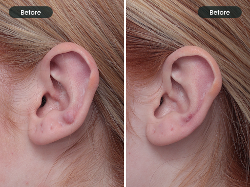
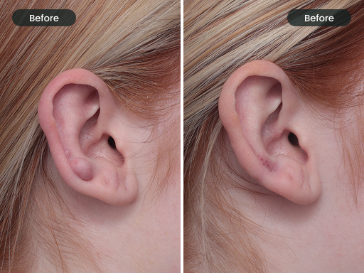
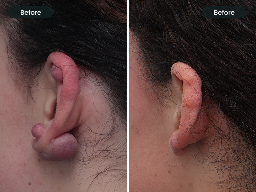
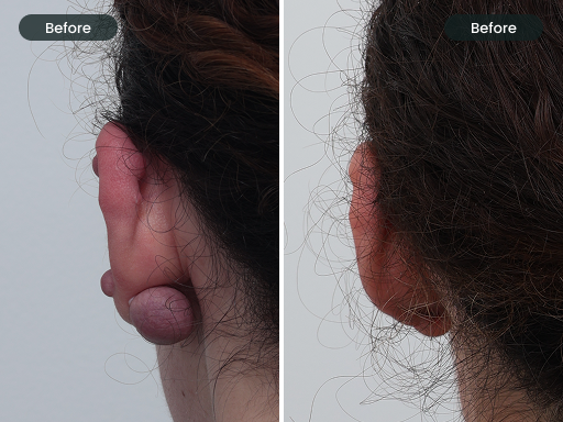
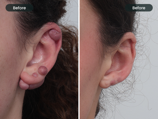
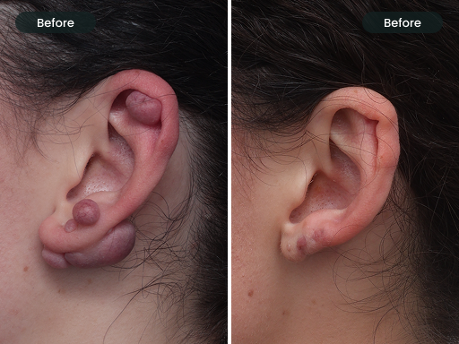
DISCLAIMER: Outcomes shown are only relevant to the individual patient and do not necessarily reflect the results other patients may experience, as results may vary due to many factors including genetics, keloid history, and adherence to aftercare. Some images may have identifiable features blurred to protect patient privacy.
Keloid vs Hypertrophic Scar — What’s the Difference?
These two types of raised scars are frequently confused, but they are biologically and clinically distinct. Getting the diagnosis right matters, because treatment approaches differ.
Hypertrophic Scar
Hypertrophic scars are raised, firm, and sometimes red — but they remain confined within the original injury borders. They often improve over 12–18 months and tend to respond well to compression and silicone therapy alone.
Keloid Scar
Keloid scars actively invade surrounding normal skin, growing beyond where the original wound was. They do not self-resolve and are driven by a persistent, dysregulated wound healing process. On the ear, true keloids frequently require a combination of surgical debulking and ongoing injection therapy to achieve lasting control.
Dr Ed assesses every patient individually to establish the correct diagnosis before any treatment is recommended.
Types & Shapes of Ear Keloids
Ear keloids are not one-size-fits-all. Their morphology — the shape, size, location, and structure — has a direct bearing on which treatment approach is most appropriate. Dr Ed classifies each keloid carefully at the initial consultation.
Pedunculated (Lobule) Keloid
The most common ear keloid. Forms a distinct, rounded, or elongated mass that hangs from the earlobe — often described as a “ball” or “mushroom” shape. Usually arises from a piercing and may be surprisingly large. Well-suited to surgical debulking given its defined base.

Sessile (Flat-Based) Keloid
A broad-based keloid that sits flush against the ear surface with no obvious neck or stalk. Often wider than it is thick. Can involve the front and back of the lobe simultaneously. Requires careful surgical planning to avoid disrupting surrounding anatomy.

Butterfly / Dumbbell Keloid
A distinctive type where keloid tissue develops on both the front and back of the earlobe, connected through the piercing tunnel. Creates a characteristic hourglass or butterfly appearance. Requires treatment of both components.

Multi-nodular Keloid
Multiple discrete keloid nodules present on the same ear, sometimes arising from different piercing sites or from separate triggers. Requires an individualised plan addressing each nodule, which may differ in size and behaviour.

Supra-auricular Keloid
Arises on the upper or outer structures of the ear (helix, anti-helix, tragus) rather than the lobe. Often associated with cartilage piercings or surgical procedures. More complex to treat due to proximity to cartilage and ear shape.

Recurrent Keloid
A keloid that has regrown following previous treatment — whether surgical, injection-based, or both. Recurrent keloids often behave more aggressively and require a more structured multimodal approach. Accurate history of prior treatments informs Dr Ed’s management plan.

The Genetics of Ear Keloids
Keloid formation is not purely random. There is a strong hereditary component — keloids run in families, and individuals with a first-degree relative who forms keloids are significantly more likely to do so themselves.
Keloids are more prevalent in individuals with darker skin phototypes (Fitzpatrick types IV–VI), with studies suggesting up to 15–20 times greater risk compared to lighter skin phototypes. However, anyone of any ethnicity or skin type can develop keloids. Asian, Hispanic, and Middle Eastern populations also show elevated keloid risk relative to Northern European backgrounds.
The genetic basis appears to involve several pathways — including dysregulation of TGF-β (transforming growth factor beta) signalling, altered collagen turnover, and abnormal apoptosis (programmed cell death) in fibroblasts. In a keloid-prone individual, the normal “stop signal” that halts collagen production after wound healing simply does not fire correctly.
Understanding your genetic risk is important — not because it changes whether treatment is possible, but because it informs the intensity of post-treatment monitoring required and the likely need for ongoing maintenance therapy.
01
Autosomal dominant tendency
Keloid susceptibility can be inherited in a dominant pattern, meaning a parent with keloids has a meaningful chance of passing on the predisposition. Siblings of affected individuals also carry elevated risk.
02
Skin phototype correlation
Darker Fitzpatrick skin types (IV–VI) are significantly overrepresented in keloid patients. This reflects differences in fibroblast activity and melanocyte biology that influence wound healing responses.
03
Age of onset matters
Keloids most commonly develop between puberty and age 30 — a period of heightened hormonal activity and robust fibroblast signalling. Post-menopausal formation is less common, and childhood keloids are relatively rare.
04
TGF-β pathway dysregulation
Overactive transforming growth factor beta signalling is a key driver. This cytokine promotes collagen synthesis; in keloid-prone individuals, its activity is poorly downregulated, resulting in continued excessive collagen deposition.
How Ear Keloids Are Treated at The DOC
Ear keloid management is not a single procedure — it is a carefully structured clinical program. Dr Ed’s approach combines surgical debulking with a planned monitoring and injection protocol, tailored to each patient’s specific keloid characteristics and history.
All procedures performed under local anaesthetic
All surgical debulking of ear keloids at The DOC is performed under local anaesthetic in the clinic setting — meaning no hospital admission, no general anaesthetic, and no overnight stay. You are awake and comfortable throughout, and can return home the same day.
1 Initial Consultation & Assessment
Dr Ed conducts a thorough assessment of your ear keloid — examining its size, morphology, location, and consistency. He will take a full history including any previous treatments, family history of keloids, your skin type, and how long the keloid has been present. This information directly shapes the treatment plan. Photography is taken for documentation and to monitor progress over time. Medicare rebates may be applicable for medically indicated consultations.
2 Surgical Debulking Under Local Anaesthetic
For most ear keloids — particularly those that are large, pedunculated, or multi-nodular — surgical debulking is the recommended first step. The procedure involves the precise excision of the keloid bulk while carefully preserving the underlying ear architecture and avoiding excessive tension on the wound edges.
The area is thoroughly anaesthetised with local anaesthetic before any incision is made, ensuring you are comfortable throughout. The technique used is carefully chosen based on the keloid’s shape and location — for example, a dumbbell keloid may require simultaneous front-and-back excision. The goal of debulking is not simply cosmetic — removing the main bulk of the keloid dramatically improves the efficacy of subsequent injection therapy.
Following excision, wound closure is planned on a case-by-case basis. Some sites are closed with fine sutures designed to minimise tension, while others — depending on their size, location, and shape — may be left to heal by secondary intention (without sutures). Dr Ed will discuss the planned closure approach with you as part of your pre-procedure consent. You will be provided with detailed aftercare instructions before you leave the clinic.
3 Post-Operative Care & Wound Monitoring
The immediate post-operative period is critical. The wound site is kept clean and protected as it heals, and Dr Ed will review you at an appropriate interval to assess healing progress. Signs of early keloid re-activity — such as the wound becoming raised, firm, or itchy — are monitored closely. Early intervention at this stage significantly improves long-term outcomes.
4 Intralesional Anti-Inflammatory Injection Program
Once the wound has reached an appropriate stage of early healing, Dr Ed commences a planned program of intralesional corticosteroid injections. These are injected directly into the scar tissue at precisely timed intervals — the timing, concentration, and frequency of which are individually calibrated based on the keloid’s size, behaviour, and your skin’s response.
The mechanism of action is well-established: corticosteroids suppress fibroblast proliferation, reduce collagen synthesis, and downregulate pro-inflammatory cytokines including TGF-β — directly targeting the biological drivers of keloid formation. Regular injection treatment also reduces keloid symptoms such as itching and tenderness.
Injections are typically spaced 4–6 weeks apart. The total number of sessions required varies considerably — simpler cases may achieve excellent control in 3–5 sessions, while larger or recurrent keloids may require 6 or more visits over an extended period. Dr Ed will give you a realistic expectation at your initial consultation based on your specific presentation.
5 Long-Term Monitoring & Maintenance
Keloids have a well-recognised tendency to recur. Even after successful treatment, ongoing monitoring is important — particularly in the first 12–24 months following the final treatment session. Dr Ed establishes a monitoring schedule appropriate to your individual risk profile. Early signs of re-activity are managed promptly to prevent re-establishment of the keloid.
Adjuvant measures — including ear compression magnets, silicone therapy, and sun protection — are incorporated into the aftercare program as complementary strategies to support long-term control. These are discussed in detail in the sections below.
Medicare Rebates May Apply
Consultations and certain keloid treatments may attract a Medicare rebate when clinically indicated. Dr Ed will assess your eligibility at your initial consultation. The DOC Clinic is a private billing clinic; fees are in line with AMA recommended specialist rates. Please call 03 9021 6022 to discuss.
The Injection Program in Detail
Intralesional corticosteroid therapy is the cornerstone of keloid management following debulking. In selected cases, Dr Ed may also utilise additional adjunctive injection agents to enhance outcomes. Understanding what to expect at each stage helps patients engage confidently with the process.
4–6
Weeks Between Sessions
Injection intervals are carefully spaced to allow the tissue to respond and recover between treatments. This timing is not arbitrary — it reflects the biology of collagen remodelling and corticosteroid pharmacokinetics in scar tissue.
3–6+
Typical Number of Sessions
Most patients require a minimum of 3 sessions, but larger or recurrent keloids may require 6 or more. Progress is reviewed at each visit and the plan adjusted accordingly. There is no fixed endpoint — treatment continues until the keloid is stable and controlled.
100%
Performed by Dr Ed Personally
All consultations and injection treatments are performed personally by Dr Ed Omarjee at both clinic locations — DOC East (Ashburton) and DOC West (Hoppers Crossing). No treatments are delegated to nurses or therapists. This ensures consistent clinical assessment and precise delivery at every visit, at both sites.
Additional Injection Agents — Beyond Corticosteroids
Adjunctive Anti-Fibrotic Therapy
An adjunctive anti-fibrotic agent may be used in low doses via intralesional injection, acting as a powerful anti-fibrotic. It works by inhibiting fibroblast proliferation — directly targeting the cells responsible for the excessive collagen production that drives keloid formation. Used at very low concentrations directly into the scar tissue, this approach is quite different in effect and dose to its broader medical uses.
Dr Ed may incorporate this adjunctive injection therapy into the post-debulking program in selected cases — particularly where a keloid has been resistant to corticosteroids alone, where recurrence has occurred following previous treatment, or where the keloid is particularly aggressive. Adjunctive therapy of this kind is often used in combination with corticosteroid injections, and evidence supports this combination as producing superior outcomes to either agent alone. The decision to use additional adjunctive therapy is made on an individual basis and discussed fully during your consultation.
Neuromodulator Adjuvant Therapy
Neuromodulator-based therapy — best known in other clinical contexts — has an emerging and well-supported role in keloid management. When injected into keloid scar tissue, it works by inhibiting TGF-β1 signalling and reducing fibroblast activity, thereby suppressing the overproduction of collagen that characterises keloid scars. It may also reduce the mechanical tension across healing wounds — a known driver of keloid formation and recurrence.
Dr Ed may utilise neuromodulator injections post-debulking in selected patients, either as a standalone adjuvant or as part of a combination injection protocol alongside corticosteroids and/or other adjunctive agents. Its use is particularly considered in areas of the ear where wound tension may be a contributing factor, and in patients where additional suppression of scar activity is clinically indicated. As with all treatment decisions, its inclusion in your program will be discussed and explained at your consultation.
What Can Patients Expect From Injections?
Each injection session involves the careful delivery of corticosteroid directly into the scar tissue using a fine needle. The ear is sensitive, and injections can be uncomfortable — though most patients find the procedure very tolerable.
Following injection, the scar may appear slightly blanched, and some temporary soreness or swelling is normal. Over subsequent weeks, patients typically notice the scar becoming softer, flatter, and less symptomatic. The colour of the keloid may also change, becoming less red or pink as vascularity reduces.
It is important to understand that results are gradual — patience and commitment to the full program are important for achieving the best possible outcome.
Potential side effects of intralesional corticosteroid therapy include localised skin atrophy (thinning of the surrounding normal skin), hypopigmentation (lightening of the skin around the injection site), and telangiectasia (small dilated blood vessels). These effects are related to dose and technique, and Dr Ed carefully titrates each injection to minimise this risk.
Rarely, patients may experience a steroid flare — a temporary increase in redness or discomfort shortly after injection. This typically settles within 24–48 hours. If you experience any unexpected changes between appointments, you are encouraged to contact the clinic.
Ear Compression Magnets — The Science
Ear compression therapy using magnetic earrings is a well-established adjunctive treatment in ear keloid management. Understanding the mechanism helps patients comply with this important part of their aftercare program.
How Compression Magnets Work
Ear compression magnets consist of two small disc magnets — one placed on each side of the earlobe — that attract each other through the tissue, applying gentle, continuous, even pressure to the surgical site or developing scar.
The scientific rationale for compression therapy in keloids is well-supported. Sustained mechanical pressure on scar tissue induces hypoxia (reduced oxygen delivery) within the developing keloid, which inhibits fibroblast proliferation and reduces collagen synthesis. Pressure also appears to induce apoptosis (programmed cell death) in the abnormal fibroblasts that drive keloid growth, while promoting a more organised collagen matrix aligned with normal skin architecture.
Magnets offer advantages over traditional compression dressings on the ear because they are discreet, easily applied and removed by the patient, and can deliver consistent pressure to the irregular contours of the earlobe without requiring complex fitting or dressings that may irritate the healing skin.
Reduces fibroblast activity through sustained mechanical pressure
Induces hypoxia within developing scar tissue, inhibiting collagen overproduction
Promotes more normal collagen fibre alignment
Discreet and easy for patient self-application
Adapts to complex earlobe anatomy better than flat compression garments
Can be worn daily alongside normal activities
Recommended Use Protocol
The effectiveness of ear compression magnets is highly dependent on consistent use. The recommended wear time is a minimum of 8 hours per day. However, some patients find this difficult due to discomfort, skin irritation, or practical lifestyle reasons. In these cases, a more conservative but still clinically meaningful period of 3–4 hours per day remains beneficial and is encouraged over no use at all. Any compression is better than none — consistency over time matters more than achieving the maximum daily target.
The recommended duration of compression therapy following ear keloid treatment is typically a minimum of 6 months, and often up to 12 months, particularly in patients with a strong keloid-forming tendency or following treatment of a recurrent keloid. Dr Ed will advise on the specific duration appropriate for your case.
Magnet strength matters — too weak and insufficient pressure is delivered; too strong and discomfort or ischaemia can occur. Dr Ed or his team will guide you on sourcing appropriately calibrated compression earrings. Purpose-designed medical pressure earrings are preferable to improvised alternatives.
Aim for a minimum of 8 hours per day; 3–4 hours per day is a practical and still effective alternative if full wear is not tolerable
Continue for 6–12 months post-treatment (as advised)
Use purpose-designed medical-grade compression earrings
Clean the site and magnets daily with gentle soap and water
Report any skin irritation, breakdown, or changes to Dr Ed promptly
Do not remove magnets for the convenience of jewellery — compliance is critical
Silicone Gel vs Silicone Sheets — Understanding the Science
Silicone therapy is one of the most evidence-supported conservative treatments for hypertrophic and keloid scars. Both silicone gel and silicone sheets are effective, but they work in complementary ways and suit different situations. Knowing which to use, and how to use it correctly, makes a significant difference to outcomes.
Silicone Gel
Topical application — dries to form a breathable film
Silicone gel is applied directly to the scar surface as a liquid that dries within a few minutes to form a thin, transparent, breathable silicone layer. It is particularly well suited to areas that are difficult to dress, including the curved contours of the ear, and to newly healed wounds where a sheet cannot be easily secured.
The mechanism involves hydration of the stratum corneum (the outermost skin layer). Scar tissue is relatively dehydrated compared to normal skin, and this low-moisture environment drives fibroblast overactivity. Silicone gel creates a semi-occlusive barrier that dramatically increases local hydration, reducing fibroblast stimulation and collagen overproduction. There is also evidence that silicone itself influences the expression of growth factors involved in fibrosis.
With consistent use over time, silicone gel has been shown to improve scar colour (reducing redness and hyperpigmentation), decrease scar thickness, improve scar elasticity and pliability, and reduce scar-associated itchiness — all of which contribute meaningfully to patient comfort and cosmetic outcome. Silicone gel is also cosmetically convenient — it can be applied under makeup or sun protection, making daily compliance easier.
Best suited for: Irregular/curved surfaces, new wounds
Application: 2× daily, thin layer
Recommended duration: Minimum 3 months; up to 6–12 months
Can wear under makeup: Yes
Compression benefit: Minimal
Silicone Sheets
Adhesive or self-adhesive film worn over the scar
Silicone sheets are self-adhesive pads of medical-grade silicone that are applied directly over the scar and worn for extended periods — typically 12–24 hours per day. They share the same hydration mechanism as silicone gel, but with an important additional benefit: they also provide a degree of gentle surface compression, which adds a mechanical component to scar modulation.
Silicone sheets are generally considered the more potent of the two formats for established, mature scars, particularly on flatter or more accessible areas. They are also reusable — a single sheet can be washed and reapplied multiple times over several weeks, making them economical for prolonged use.
On the ear, sheets can be challenging to apply to the earlobe’s curved surface, and may lift or peel with movement. This is one reason silicone gel is often preferred for the active treatment phase on the ear itself, with sheets reserved for accessible scar margins or post-auricular areas where they can lie flat.
Best suited for: Established scars, accessible flat areas
Application: 12–24 hours/day, continuous wear
Recommended duration: Minimum 3 months; up to 12 months
Can wear under makeup: Yes — wash and reapply
Compression benefit: Moderate (surface pressure)
Why Duration Matters — The Collagen Remodelling Timeline
Scar maturation is a long biological process. Active collagen remodelling in a healing scar continues for up to 2 years after the initial wound. The recommendation for a minimum of 3 months of continuous silicone therapy reflects the time required to meaningfully influence collagen organisation during this remodelling window.
Studies consistently show that compliance is the single most important predictor of silicone therapy effectiveness. Sporadic use produces significantly worse outcomes than daily, consistent application. Dr Ed recommends incorporating silicone therapy into a daily skincare routine — applying it at the same time each day to build a reliable habit throughout the treatment course.
Before & After Gallery — Ear Keloids
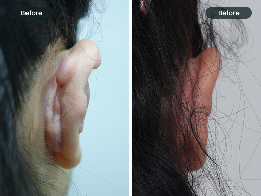
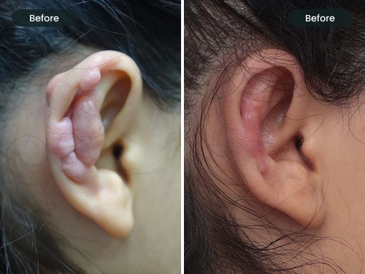
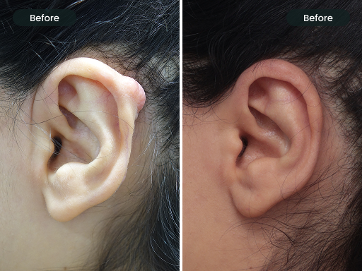

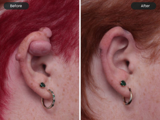
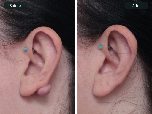
DISCLAIMER: Outcomes shown are only relevant to the individual patient and do not necessarily reflect the results other patients may experience, as results may vary due to many factors including genetics, keloid history, and adherence to aftercare. Some images may have identifiable features blurred to protect patient privacy.
Risks & Expected Results
All medical procedures carry risk. Being fully informed before proceeding is something Dr Ed takes seriously, and a comprehensive discussion of risks and realistic outcomes forms part of every consultation.
Recurrence
Keloid recurrence is the most significant risk. Even with optimal treatment, keloids can regrow — which is why post-treatment monitoring and adjuvant therapy are critical components of the program.
Infection
Any surgical procedure carries a small risk of infection. Careful wound care instructions and prompt review if concerns arise minimise this risk.
Skin Atrophy
Repeated intralesional corticosteroid injections can cause thinning of the surrounding normal skin or the development of small blood vessels (telangiectasia). Careful dosing minimises this risk.
Hypopigmentation
Lightening of the skin colour around the injection site is a recognised side effect of intralesional steroids, particularly relevant in darker skin phototypes.
Scarring
All excision procedures leave some form of scar. In keloid-prone individuals, any new wound has the potential to form another keloid — which is why the post-operative injection program is so important.
Need for Further Treatment
Keloid management is an ongoing process. Additional treatment sessions beyond the initial plan may be required depending on how the scar responds over time.
Post-Inflammatory Hyperpigmentation (PIH)
PIH is a darkening of the skin at or around the treatment site, caused by increased melanin production triggered by inflammation or injury. It is more common in darker skin phototypes (Fitzpatrick IV–VI) and can occur following both surgical debulking and injection therapy. PIH is not permanent — it typically fades gradually over weeks to months — but diligent sun protection of the treated area is important to prevent it deepening. Dr Ed will advise on appropriate management if PIH develops following your treatment.
Keloid Recurrence — What You Need to Know
Recurrence is the defining challenge of keloid management, and the risk that distinguishes keloid scars from almost every other scar type. Even after successful treatment, keloids can regrow in susceptible individuals — and the likelihood of this is not the same for everyone. Recurrence risk is shaped by a combination of individual patient factors and the specific characteristics of the keloid itself.
What the Evidence Says: Surgery Alone vs Structured Post-Treatment Care
The published medical literature is unambiguous on this point. Surgical excision of ear keloids without any follow-up treatment carries a recurrence rate of 45–100%. In many cases, the recurrent keloid grows back larger and more aggressive than the original. This is why surgery alone is not considered an appropriate treatment for keloids — it is the starting point, not the solution.
When surgical debulking is followed by a structured, monitored program of intralesional corticosteroid injections, the picture changes substantially. Studies report recurrence rates as low as 5-20% for earlobe keloids treated with combined excision and post-operative intralesional corticosteroid therapy — a dramatic improvement over surgery alone. The evidence strongly supports the combination of debulking and a bespoke, prolonged injection program as the most effective non-radiotherapy approach to ear keloid management.
45–100%
Surgery alone
Recurrence rate after surgical excision of ear keloids without any follow-up treatment.
5-20%
Excision + structured injection program
Recurrence rate reported for earlobe keloids treated with combined excision and post-operative intralesional corticosteroid therapy.
Factors That Influence Recurrence Risk
Not all ear keloids carry the same recurrence risk. Dr Ed takes all of the following into account when planning the duration and intensity of post-treatment monitoring for each patient:
Age of the Keloid
Younger, more recently formed keloids tend to be more biologically active — fibroblasts are still in a heightened state of collagen production. Older, long-standing keloids are generally more quiescent. A keloid that has been present and growing for only 6–12 months is typically at higher recurrence risk than one that has been stable and unchanged for several years.
Symptomatic Keloids
A keloid that is actively itchy, tender, or painful is almost always a sign that it is biologically active — inflammation is ongoing within the scar tissue, fibroblast activity is elevated, and the keloid is likely still growing. Symptomatic keloids carry a significantly higher recurrence risk than asymptomatic ones and typically require a more intensive and prolonged post-treatment program.
Size of the Keloid
Larger keloids contain a greater volume of abnormal fibroblasts and collagen — and leave a correspondingly larger wound bed following debulking. Larger maximum lesion diameter is a well-documented independent risk factor for keloid recurrence in the medical literature. Very large keloids may require more injection sessions over a longer period to achieve durable control.
Number of Keloids
Patients with multiple keloids — whether on the same ear or across multiple sites — often have a stronger underlying keloid-forming tendency than those with a single lesion. Multiple lesions reflect a more systemic predisposition, and each individual keloid may require its own treatment plan and monitoring schedule.
Shape and Morphology of the Keloid
Keloid shape has a meaningful bearing on recurrence risk — and this is a nuance that is often overlooked. Pedunculated keloids (those with a defined stalk or neck) generally carry a lower recurrence risk following surgical debulking, because their base of attachment is narrower, the wound bed is more defined, and wound tension after excision is typically lower. Broad-based (sessile) keloids — which spread widely across the earlobe without a clear neck — are more challenging: they involve a larger wound area, healing is under greater tension, and the recurrence risk is correspondingly higher. Dumbbell or butterfly-shaped keloids — which pass through the piercing tunnel and involve both the front and back of the earlobe — are among the most complex morphologically, requiring careful treatment of both components and carrying a higher recurrence risk due to the anatomical tension and the extent of tissue involved. Dr Ed classifies every keloid by morphology at the initial consultation, and this directly informs the intensity and duration of the post-treatment program.
The Honest Expectation
For keloid-prone individuals, ongoing vigilance is a long-term commitment. Dr Ed establishes a monitoring schedule following active treatment — with more frequent reviews in the first 12 months, tapering as the scar remains stable. Early signs of re-activation — such as itching, firmness, or slight re-elevation — are acted upon promptly. In many cases, a short course of early re-treatment can prevent a full recurrence from re-establishing. The goal is durable control, and for the vast majority of patients who remain engaged with monitoring and timely re-treatment, this is entirely achievable.
What Are Realistic Expected Results?
Significant size reduction
Most patients achieve substantial flattening and reduction in keloid bulk following debulking and injection therapy — often 70–90% improvement in volume in compliant patients.
Symptom relief
Itching, tenderness, and the sensation of tension in the keloid are usually among the earliest benefits patients notice following the commencement of injection therapy.
Long-term control, not cure
In keloid-prone individuals, the goal of treatment is sustained control rather than a permanent cure. With monitoring and maintenance, most patients achieve durable, excellent results.
Why Melbourne Patients Choose Dr Ed for Ear Keloid Treatment
20+ Years of Dedicated Keloid Experience
Dr Ed has been treating keloid scars since 2006 and has built one of Melbourne’s most extensive keloid treatment practices, seeing patients from Hoppers Crossing, Werribee, Point Cook, Ashburton, Camberwell, Glen Iris, and across metropolitan Melbourne. He has personally treated over 1,000 ear keloid patients — providing a depth of dedicated clinical experience that directly benefits every new patient.
Every Consultation & Treatment Performed by Dr Ed Personally
At The DOC, all consultations and treatments are conducted by Dr Ed himself. No nurses. No therapists. The person who assesses you is the same person who treats you — ensuring clinical continuity at every stage of your care.
Structured, Evidence-Based Treatment Programs
Ear keloid management at The DOC follows an evidence-guided, multimodal approach — combining surgical precision with systematic injection therapy and adjuvant measures. Each plan is individually tailored, not generic.
Two Convenient Melbourne Locations
With clinics in Hoppers Crossing (DOC West, Melbourne’s West) and Ashburton (DOC East, Melbourne’s Inner East), Dr Ed provides ear keloid treatment to patients across Melbourne — including Werribee, Point Cook, Laverton, Williams Landing, Altona, Wyndham Vale, Williamstown, Footscray, Geelong, Ballarat, and Bendigo from Hoppers Crossing, and Camberwell, Glen Iris, Malvern, Hawthorn, Glen Waverley, Box Hill, Ashwood, Carnegie, and surrounds from Ashburton. No GP referral is required.
CPCA & ACAM Certified — Medically Trained Cosmetic Physician
Dr Ed holds fellowship certification with the Cosmetic Physicians College of Australasia (CPCA) and the Australasian College of Aesthetic Medicine (ACAM), reflecting his commitment to ongoing medical education and evidence-based practice.
Holistic Aftercare Support
Treatment doesn’t end when you leave the clinic. Dr Ed provides detailed, personalised aftercare advice covering wound care, silicone therapy, compression magnet use, sun protection, and monitoring schedules — giving you the best possible foundation for long-term success.
Frequently Asked Questions — Ear Keloids
Can I get an ear keloid from a normal lobe piercing?
Yes, absolutely. Even a standard, professionally performed earlobe piercing can trigger an ear keloid in a susceptible individual. This is why ear keloids are among the most commonly encountered in clinical practice. If you have a personal or family history of keloid formation, you should be aware of this risk before having any ear piercing performed. Those with a known keloid tendency may wish to consult Dr Ed before piercing, or to seek early review if any raised tissue develops around a piercing site.
Will the keloid come back after treatment?
Recurrence is the central challenge in keloid management, and the honest answer is that keloids do carry a meaningful recurrence risk — particularly when treated with surgery alone. This is precisely why Dr Ed’s approach combines surgical debulking with a carefully planned, sustained injection program and adjuvant compression/silicone therapy. Studies consistently show that this multimodal approach achieves significantly lower recurrence rates than any single modality alone. Long-term monitoring following treatment completion also allows early re-treatment if any signs of regrowth appear.
Does ear keloid treatment hurt?
Surgical debulking is performed under local anaesthetic, so you should not feel pain during the procedure itself — though you will feel pressure and awareness of movement. The injection of local anaesthetic itself produces a brief stinging sensation as it is administered. Intralesional steroid injections can be uncomfortable, as the tissue is being injected under pressure. Dr Ed uses techniques to minimise discomfort at each visit, and most patients find the injections very manageable in a clinical setting. If you have concerns about pain, please discuss this with Dr Ed at your consultation.
Do I need a GP referral to see Dr Ed about my ear keloid?
No GP referral is required to book a consultation at The DOC Clinic. You can book directly online or by calling the clinic on 03 9021 6022.
How long does the full ear keloid treatment program take?
This depends significantly on the size, behaviour, and history of your keloid. A straightforward, first-time ear lobe keloid may be well-controlled within 4–6 months from the initial debulking procedure through to the completion of injection therapy. More complex, large, or recurrent keloids may require an active treatment period of 9–18 months, followed by an ongoing monitoring phase. Adjuvant silicone therapy and compression magnet use are recommended for a minimum of 6–12 months regardless of the size of the keloid. Dr Ed will provide a realistic individualised timeline at your consultation.
Can I get my ear re-pierced after keloid treatment?
Re-piercing after keloid treatment is generally not recommended. Any new wound to the ear — including re-piercing — carries a significant risk of forming another keloid in an already keloid-prone individual. If re-piercing is important to you, this should be discussed in detail with Dr Ed, who can advise on timing, risk-reduction strategies (such as prophylactic injection at the time of piercing), and whether your specific case makes re-piercing a reasonable consideration.
Are compression magnets safe? Do they cause any problems?
Medical-grade ear compression magnets are considered safe for most patients. The magnetic field they generate is localised and very low-level. They should not be used by patients with pacemakers or certain metal implants — Dr Ed will screen for contraindications at your consultation. Potential issues include skin irritation or pressure sores if applied too tightly, or if worn continuously without daily hygiene care. Dr Ed will guide you on appropriate application technique and hygiene to avoid these problems.
Does Medicare cover ear keloid treatment?
Medicare rebates may be applicable for consultations and certain medically indicated keloid treatments. Purely cosmetic consultations are not Medicare rebatable. Dr Ed will assess your eligibility at your initial consultation. The DOC Clinic is a private billing clinic, and fees are charged in line with AMA recommended specialist rates. Full payment is required on the day. Please call the clinic to discuss fee arrangements in advance if you would like further guidance.
How is silicone gel different from silicone sheets — which should I use?
Both are effective and share the same primary mechanism — hydration of the scar surface to reduce fibroblast overactivity. Silicone gel is generally preferred on the ear because it conforms to curved surfaces, dries invisibly, and can be applied under other products. Silicone sheets provide a small additional compression effect and are highly effective on flatter or more accessible scars. For ear keloid management, Dr Ed typically recommends silicone gel as the primary silicone-based adjuvant, with sheets potentially used on more accessible scar margins or post-auricular areas. The most important factor is consistent daily use for the recommended duration — at least 3 months, and ideally 6–12 months.
I had a keloid treated elsewhere and it came back. Can Dr Ed still help?
Yes. Recurrent keloids are a significant part of Dr Ed’s practice. A detailed history of your previous treatment — including what was done, when, and how the keloid responded — is important information that will inform the new management plan. Recurrent keloids can often still be effectively managed, though they may require a more intensive or prolonged treatment course. Please bring any relevant documentation or photographs from your previous treatment to your consultation if available.
Book Your Ear Keloid Consultation in Melbourne
Ear keloid consultations and treatment are available at two Melbourne locations: The DOC West in Hoppers Crossing (serving Melbourne’s western suburbs and beyond, including Werribee, Point Cook, Laverton, Williams Landing, Altona, Wyndham Vale, Williamstown, Footscray, Geelong, Ballarat, and Bendigo) and The DOC East in Ashburton (serving Melbourne’s inner east including Camberwell, Glen Iris, Malvern, Hawthorn, Glen Waverley, Box Hill, Ashwood, and Carnegie). No GP referral is required.
DOC West
302 Heaths Road, Hoppers Crossing VIC 3029
DOC East
47A Karnak Rd, Ashburton VIC 3147
Serving patients across Melbourne including: Hoppers Crossing, Werribee, Point Cook, Laverton, Williams Landing, Altona, Wyndham Vale, Truganina, Tarneit, Williamstown, Footscray, Geelong, Ballarat, Bendigo (from DOC West) and Ashburton, Camberwell, Glen Iris, Malvern, Hawthorn, Glen Waverley, Box Hill, Ashwood, Carnegie (from DOC East).
MED0001186584 | As with any medical procedure, individual results may vary. All procedures carry risks — discussed in full at your consultation.


